Echocardiogram
An echo provides important details for children with heart symptoms or known heart conditions that help doctors decide on the best care plan.
What to Expect
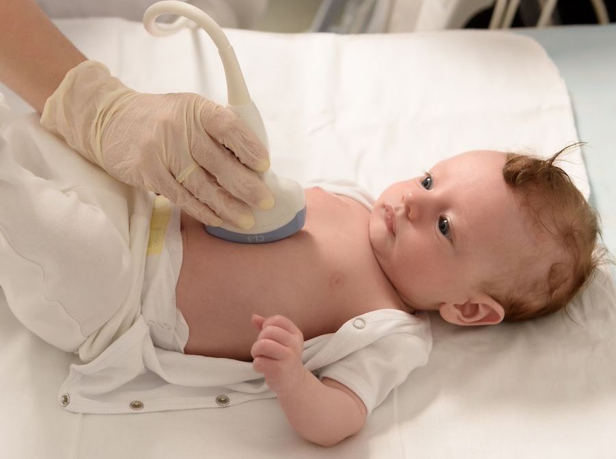
During the test, your child will lie comfortably on an exam table, usually on their left side. A technician will put a small amount of gel on your child’s chest and use a device called a transducer to get “pictures” of the heart. The gel helps get clear images, and the transducer moves gently across the chest. The whole process usually takes 30 minutes to an hour and is completely painless.
How to Prepare Your Child
- Explain the Process: Let your child know it’s like taking a picture of their heart. You can describe the gel as feeling a bit cool and the device as smooth and gentle.
- Dress Comfortably: Loose clothing makes it easier for the technician to position the transducer on their chest.
- Bring a Comfort Item: If your child has a favourite toy or blanket, bringing it can help them feel more at ease.
Benefits of Echocardiogram
Echocardiograms offer several important benefits, including:
- Non-Invasive: It is a painless procedure that does not require incisions or radiation exposure.
- Real-Time Heart Function Monitoring: It provides immediate results on how the heart muscles and valves function.
- Detailed Visualisation: Helps detect abnormalities in heart structure, such as enlarged chambers, weakened heart muscles, or valve dysfunction.
- Early Diagnosis: Assists in the early detection of heart conditions, enabling timely treatment.
- Guidance for Treatment: Offers valuable insights for doctors to plan or adjust treatment strategies for cardiac patients.
- Monitoring Progress: Useful for tracking the effectiveness of treatments or surgeries over time.
Types of Echocardiograms
There are several types of echocardiograms, each designed for specific diagnostic purposes:
- Transthoracic Echocardiogram (TTE): The most common type, where a transducer is placed on the chest to capture heart images through the chest wall. It is simple, non-invasive, and provides a comprehensive view of the heart’s structure and function.
- Transesophageal Echocardiogram (TEE): In this type, a specialised probe is inserted down the oesophagus to capture clearer images of the heart, as it lies closer to the organ than the chest wall. This is especially useful for assessing hard-to-view areas and more detailed examinations.
- Doppler Echocardiogram: This procedure observes blood flow through the heart’s chambers and valves. It can detect abnormal blood flow and help diagnose issues like valve stenosis or regurgitation.
- Stress Echocardiogram: Performed during or after exercise to see how the heart functions under physical stress. This test helps assess for conditions like coronary artery disease.
- 3D Echocardiogram: This technique offers three-dimensional imaging to better visualise heart defects and structures. It is particularly useful for complex heart surgeries and valve assessments.
Alternative Options to Echocardiogram
While echocardiograms are highly effective, there are alternative tests that might be used based on specific patient needs:
- Electrocardiogram (ECG/EKG): Measures the heart's electrical activity. It’s often used to detect arrhythmias but doesn’t provide detailed images of heart structures.
- Cardiac MRI: Uses magnetic fields and radio waves to create detailed images of the heart and surrounding structures. It is useful for assessing complex congenital heart defects and tissue damage after a heart attack.
- CT Coronary Angiography: A non-invasive imaging method using X-rays to visualise the heart’s blood vessels. It helps identify blockages or narrowing in the coronary arteries.
- Nuclear Cardiology (e.g., SPECT or PET scans) involves using a small amount of radioactive material and a scanning device to check blood flow to the heart muscle. It can provide functional information on heart perfusion and viability.
- Chest X-ray: Though not as detailed as an echocardiogram, it can show the size of the heart and detect fluid buildup in the lungs.
- Cardiac Catheterization is an invasive test in which a thin tube (catheter) is inserted into a blood vessel to reach the heart. It’s primarily used to visualise and assess blood flow and pressure within the heart chambers and often includes angiography.
Preparation Before an Echocardiogram
Preparing for an echocardiogram is generally straightforward. Here are some common guidelines:
- Wear comfortable, loose clothing. You may be asked to change into a hospital gown for the test.
- Most echocardiograms have no special dietary restrictions, so you can eat and drink as usual. However, if you are undergoing a stress echocardiogram or a transesophageal echocardiogram (TEE), you might be instructed to avoid food and drink for several hours before the test.
- Inform your doctor about any medications or supplements you are taking. You may need to adjust or stop certain medicines before the test, especially for a stress echocardiogram.
- If a stress echocardiogram is planned, your doctor might advise you to avoid caffeine or certain stimulants, as these can affect your heart rate and the test results.
- If you have a TEE, you may be sedated during the procedure, so it's advisable to arrange for someone to drive you home afterwards.
What Happens During an Echocardiogram?
The process of an echocardiogram varies slightly depending on the type, but a standard transthoracic echocardiogram (TTE) typically involves:
- Preparation: You will lie on an examination table, usually on your left side. The technician or doctor will apply a gel to your chest to help the transducer make better contact with the skin and produce more precise images.
- Imaging: The transducer, a handheld device that emits sound waves, will be moved around different parts of your chest. The sound waves bounce off the heart structures and create echoes, which are then converted into moving images of the heart on a monitor. You may be asked to change positions or hold your breath briefly to obtain better images.
- Duration: The entire procedure generally takes between 30 minutes to an hour. It is painless and non-invasive.
A transesophageal echocardiogram (TEE) involves using a sedative and inserting a thin tube with a transducer down the throat for closer and clearer imaging. A stress echocardiogram involves monitoring the heart during physical activity, typically on a treadmill or stationary bike or with medication that simulates exercise.
What Happens After the Test?
The test is done immediately after the images are taken, and your child can return to their usual activities immediately. The doctor will review the images and discuss any findings with you, usually within a few days. If there are any next steps, your doctor will explain them to you.
Why an Echocardiogram Matters
An echocardiogram provides a detailed picture of your child’s heart, helping doctors make the best decisions about their heart health and ongoing care.
What Are the Risks of an Echocardiogram?
Echocardiograms are generally very safe, especially the standard type used most often for children. However, depending on the type of echocardiogram, there are a few possible risks or side effects to be aware of:
Standard Transthoracic Echocardiogram (TTE)
- Minimal Risk: This type of echocardiogram is non-invasive and painless. The transducer, or probe, may cause mild pressure on the chest, which could feel slightly uncomfortable for some children, but there is no pain or lasting discomfort.
- No Radiation or Sedation: This test is very low-risk because it does not involve exposure to radiation or the need for sedation.
Transesophageal Echocardiogram (TEE)
- Sedation Reactions: Since a TEE involves a small probe placed in the throat to get clearer heart images, mild sedation or anesthesia is often used. Some children may experience drowsiness after sedation, so they’ll need time to recover.
- Sore Throat: Afterward, children may feel a mild sore throat, which usually goes away within a few hours.
- Rare Risks: In rare cases, minor injury to the mouth or throat or an unexpected reaction to the sedation could occur.
Stress Echocardiogram
- Risk of Dizziness or Shortness of Breath: A stress echo is done while your child exercises or with medicine to simulate exercise. Some children may experience mild dizziness, shortness of breath, or slight discomfort due to the physical activity.
- Very Rare Complications: In rare cases, exercise might trigger abnormal heart rhythms or other symptoms, but these tests are done with close supervision to manage any issues.
Overall Safety
Echocardiograms are considered very safe and low-risk, especially the standard TTE, the most commonly used test for children. If your child’s doctor recommends a TEE or stress echo, they’ll take extra care to minimise risks and ensure your child’s comfort throughout the process.
What if Echocardiogram is Delayed?
Delaying an echocardiogram can have varying implications based on individual health circumstances:
- Delayed Diagnosis: Postponing the test might delay identifying heart conditions, potentially allowing diseases to progress unchecked.
- Progression of Symptoms: Without timely assessment, symptoms such as shortness of breath, chest pain, or fatigue may worsen, affecting quality of life.
- Impact on Treatment: Early detection often leads to more effective treatment. Delays can limit therapeutic options and may result in less favourable outcomes.
If an echocardiogram is recommended, it's advisable to schedule it promptly to ensure optimal heart health management.